
Lymphangio-myomatosis : Microscopic

slide 13 of 20


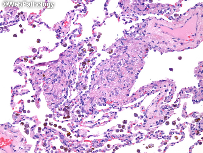
Comments:
Lymphangiomyomatosis in Lungs. High magnification view showing proliferation of plump spindle-shaped myoid cells arranged in short fascicles causing thickening of the alveolar septal wall. The spindle cells have eosinophilic cytoplasm and uniform nuclei lacking cytologic atypia or increased mitotic activity. Aggregates of lymphocytes may be seen between the myoid cells. There may be type II pneumocyte hyperplasia, especially in individuals with tuberous sclerosis.
slide 13 of 20