
Liver Hemangioma : Gross Pathology


Comments:
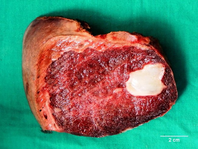
Gross Pathology - Cavernous Hemangiomas of Liver: They are larger, usually solitary, reddish-brown, well-circumscribed, unencapsulated masses with a honeycombed surface. Some may be pedunculated. The location may be subcapsular (more common) or deep parenchymal. Lesions larger than 5 cm are arbitrarily referred to as giant cavernous hemangiomas. Some may reach 20-30 cm in size. Smaller satellite lesions may be present adjacent to the main tumor mass. This 9 cm cavernous hemangioma was resected from the liver of a 42 y/o female who presented with 1-yr history of intermittent right upper quadrant pain. The whitish area on the right was composed of dense hyalinized collagenous tissue indicating chronicity of the lesion. Image courtesy of: Dr. Sanjay D. Deshmukh, Professor of Pathology, Pad. Dr. Vithalrao Vikhepatil Medical College, Ahmednagar, INDIA.