
Gout : Radiographic Features

slide 10 of 42


Comments:
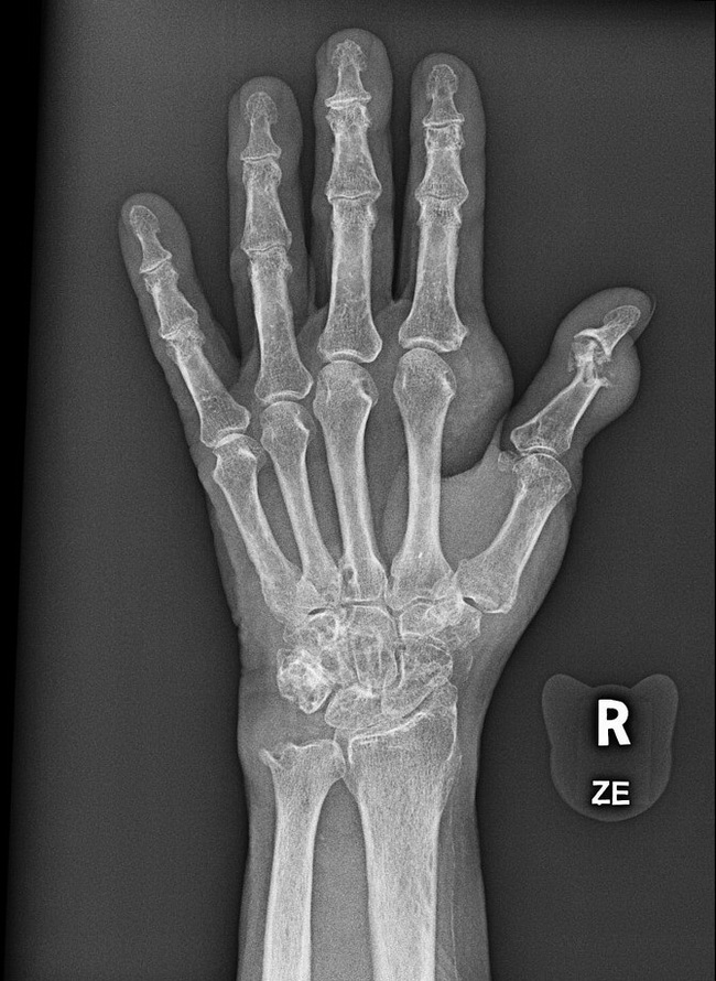
80 y/o male with persistent wrist pain and tenderness over left ulnar bone. The radiograph shows multiple well-circumscribed punched-out bony erosions with sclerotic margins, particularly in the ulnar styloid, triquetrum and distal part of first proximal phalanx. There are juxta-articular soft tissue cloudy densities and swelling distal to the ulnar and radial to the second metacarpo-phalangeal (MCP) joint. Case courtesy of Dr Matthew Lukies, Radiopaedia.org. From the case rID: 53874
slide 10 of 42