
Ameloblastic Fibroma
slide 1 of 8


Comments:
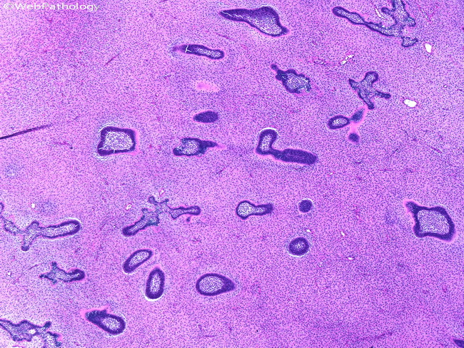
Ameloblastic fibroma usually involves the mandible (bicuspid-molar region) of young individuals. This case is from a 12 y/o male who presented with a well-circumscribed, radiolucent mass in the right mandible. Grossly, the resected specimen had pink-tan whorled appearance with gelatinous consistency. Low-power view shows nests of ameloblastic cells in a fibroblastic stroma.
slide 1 of 8