
Chondroblastoma
slide 1 of 15


Comments:
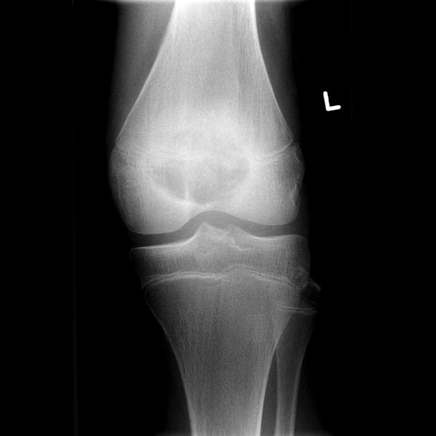
This plain film of the left knee joint is from a 14 y/o male who presented with h/o pain. A sharply defined oval lucent lesion is centered on the epiphysis of the distal femur, and appears to transgress the growth plate (which remains open). It has a narrow zone of transition and no convincing matrix calcification. A joint effusion is present. No periosteal reaction is present. This case demonstrates typical appearances of a chondroblastoma, in a typical location. The lesion was curetted and the diagnosis was confirmed on histolopathologic examination. Case produced with permission, courtesy of Dr. Frank Gaillard. Radiopaedia. Complete case is here.
slide 1 of 15