
Hydatid Cyst : Kidney

slide 9 of 32


Comments:
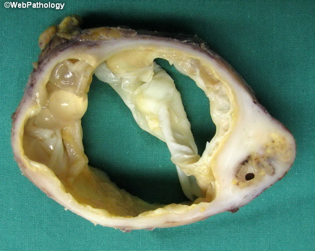
Kidney is one of the less frequently involved organs in Echinococcosis (Hydatid cyst). The photograph shows a cross section through a large hydatid cyst in the kidney with complete destruction of the native renal architecture. A few daughter cysts can be seen along the left side of the cyst. The cyst lumen is traversed by folded membranous structures. The membranes have a laminated structure histologically and are lined by germinal epithelium which gives rise to several daughter cysts (Brood capsules) inside which grow scoleces. Case courtesy of : Dr. Abdul Haleem; used with permission.
slide 9 of 32