
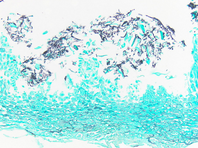
Candida Esophagitis : GMS Stain

slide 14 of 20


Comments:
Candida Esophagitis: GMS stain highlights pseudohyphae and yeast forms in this esophageal biopsy. Image courtesy of: Dr. Phoenix Bell
slide 14 of 20