
Ependymoma

slide 5 of 48


Comments:
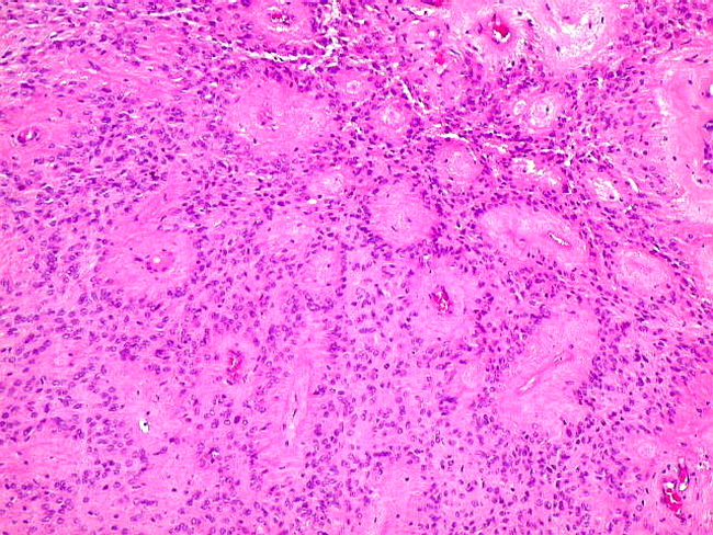
Classic ependymoma is a well-circumscribed and a cellular neoplasm with sheet-like growth pattern. The tumor cells are frequently arranged around blood vessels creating perivascular pseudorosettes, many of which can be seen in this image. Approximately 90% of ependymomas involve the brain, most of them are infratentorial. Most of these cases involve children with the peak incidence in the first decade. The remaining 10% involve the spinal cord, mostly in adults. Courtesy of: Dr. Luciano de Souza Queiroz, Dept. of Pathology, Faculty of Medical Sciences, State University of Campinas (UNICAMP), Campinas, Sao Paulo State, BRAZIL. Additional images are here.
slide 5 of 48