
Sebaceoma


Comments:
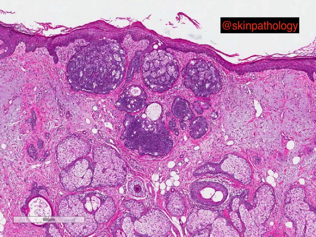
Sebaceoma consists of variably-sized lobules scattered in the dermis, often with the involvement of the overlying epidermis. The lobules lack an organized architecture and are composed of a random mixture of abundant basaloid cells and small numbers of mature sebocytes. DIFFERENTIAL DIAGNOSIS: Sebaceous adenoma: The lobules in sebaceous adenoma try to recapitulate the normal architecture of mature sebaceous glands and show basaloid germinative cells in the periphery and pale-staining mature sebocytes in the center. Sebaceous carcinoma: Most sebaceous carcinomas have significant nuclear atypia, prominent nucleoli, increased mitotic activity, and infiltrating margins. Well-differentiated examples may mimic sebaceoma. Basal cell carcinoma (BCC) with sebaceous differentiation: Sebaceous differentiation is sometimes an incidental finding in a BCC. The presence of peripheral palisading, cleft formation, and cytologic features are usually sufficient in arriving at the correct diagnosis. In challenging cases, immunostains can be helpful. EMA and D2-40 are positive in sebaceomas whereas BerEP4 is positive in BCC. Image courtesy of: Paul Drury, MD, Perth, Western Australia.