
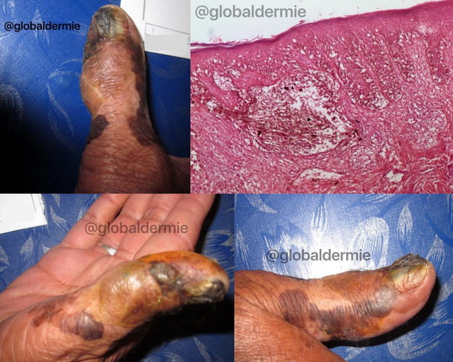
Acral Lentiginous Melanoma


Comments:
Case History: 80 y/o male patient from a developing country in Asia with a 2 yr history of pigmented lesions on his thumb. Diagnosis: Acral Lentiginous Melanoma (ALM). The patient presented at Stage 4 with liver metastases. Discussion: ALM is the most common type of melanoma in dark-skinned and Asian populations. This is because the frequency of other types of melanoma is low in these patients, not because the incidence of ALM is any higher than in white people. Median age at diagnosis is 50 years. The thumb and big toe are more frequently involved than other digits. ALMs demonstrate a junctional growth pattern and have indistinct margins. The lesion often extends laterally far beyond the clinically apparent margin. Over time, a vertical (invasive) growth phase develops. Periungual hyperpigmentation and Hutchinson's sign (dark pigment under the nail extending to involve the adjacent nail fold) is an ominous sign suggesting melanoma in the nail matrix. The early changes of ALM may be light brown and uniformly pigmented. Over time, the lesion becomes darker and nodular and may ulcerate. Metastases to the epitrochlear and axillary nodes are common, because the diagnosis is often delayed. Subungual melanoma may be misdiagnosed as onychomycosis, verruca vulgaris, chronic paronychia, subungual hyperkeratosis, etc. Images and comment courtesy of: Josette McMichael, MD, Adj Asst. Professor, Emory University, Atlanta, GA, USA. Used with permission.