
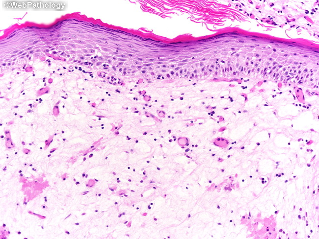
Keratinizing Squamous Metaplasia
Home
 Genitourinary
Urinary Bladder
Squamous Lesions in Urinary Bladder
Keratinizing Squamous Metaplasia
Genitourinary
Urinary Bladder
Squamous Lesions in Urinary Bladder
Keratinizing Squamous Metaplasia

slide 3 of 18


Comments:
This bladder biopsy is from a 78 y/o male with history of repeated urinary tract infections and urinary obstruction. Cystoscopy showed a whitish patch on the anterior bladder wall which was biopsied. Sections revealed keratinizing squamous metaplasia with hyperkeratosis and a granular cell layer. Keratinizing squamous metaplasia is a risk factor for development of squamous cell carcinoma and requires close follow-up.
slide 3 of 18