
Complete Hydatidiform Mole
Home
 Gynecologic
Placenta & Trophoblastic Lesions
Complete Hydatidiform Mole
Complete Hydatidiform Mole
Gynecologic
Placenta & Trophoblastic Lesions
Complete Hydatidiform Mole
Complete Hydatidiform Mole
slide 1 of 8


Comments:
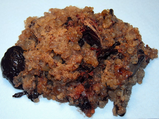
Complete hydatidiform mole almost always lacks an embryo/fetus. Before the widespread use of pelvic ultrasound during pregnancies, complete mole usually presented between 11 and 25 weeks with vaginal bleeding and/or excessive uterine enlargement for gestational age. The hCG levels are usually markedly elevated. Grossly, a complete mole consists of variably sized swollen vesicular villi. Image courtesy of Steven O'Connor, MD, Houston, Texas. Used with permission
slide 1 of 8