
Glandular Differentiation in Urothelial Carcinoma
Home
 Genitourinary
Urinary Bladder
Glandular Lesions in Urinary Bladder
Glandular Differentiation in Urothelial Carcinoma
Genitourinary
Urinary Bladder
Glandular Lesions in Urinary Bladder
Glandular Differentiation in Urothelial Carcinoma

slide 39 of 50


Comments:
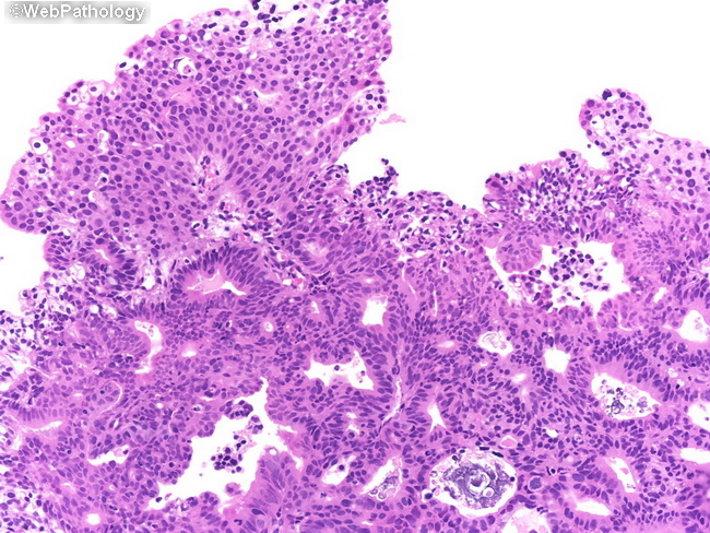
The image shows conventional urothelial carcinoma in the upper part with areas of glandular differentiation underneath. This feature is seen in about 10% of urothelial carcinomas. It is not considered to be of any clinical significance. However, it is recommended that such features be noted in the pathology report. This information may be helpful if the tumor metastasizes.
slide 39 of 50