
Mucinous Borderline Tumor

slide 36 of 84


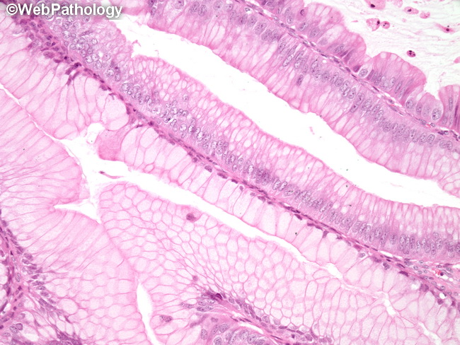
Comments:
Cytologic atypia in a mucinous borderline tumor - intestinal type: Juxtaposition of benign areas (lower left) and borderline areas (upper right) in a mucinous borderline tumor. The benign areas are lined by a single layer of mucinous epithelium with basally-located hyperchromatic nuclei lacking atypia. The borderline areas show nuclear enlargement and hyperchromasia with prominent nucleoli. They resemble adenomatous colonic polyp. High-grade nuclear features are not present.
slide 36 of 84