
Amyloidosis of Bladder

slide 32 of 39


Comments:
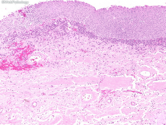
This bladder biopsy is from a 70 y/o male with left flank pain who was found to have left hydronephrosis on imaging studies. Cystoscopy showed a mass partially obstructing the left ureteral orifice which was biopsied. The lamina propria has deposits of amorphous eosinophilic material suggestive of amyloid. With Congo Red stain, these foci had the characteristic apple-green birefringence under polarized light confirming the diagnosis.
slide 32 of 39