
Schwannoma

slide 4 of 62


Comments:
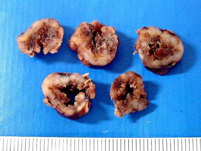
Schwannoma of the cauda equina region of spinal cord in a 30 y/o male. The tumor is multinodular and appears encapsulated. Serial sectioning showed foci of hemorrhage and cystic change. Secondary changes are more likely to be seen in the larger deep-seated tumors arising in the retroperitoneum or posterior mediastinum. Courtesy of: Dr. Luciano de Souza Queiroz, Dept. of Pathology, Faculty of Medical Sciences, State University of Campinas (UNICAMP), Campinas, Sao Paulo State, BRAZIL. Additional images are here.
slide 4 of 62