
Papillary Endothelial Hyperplasia
Home
 Soft Tissue
Vascular & Lymphatic
Papillary Endothelial Hyperplasia
Papillary Endothelial Hyperplasia
Soft Tissue
Vascular & Lymphatic
Papillary Endothelial Hyperplasia
Papillary Endothelial Hyperplasia

slide 3 of 3

Comments:
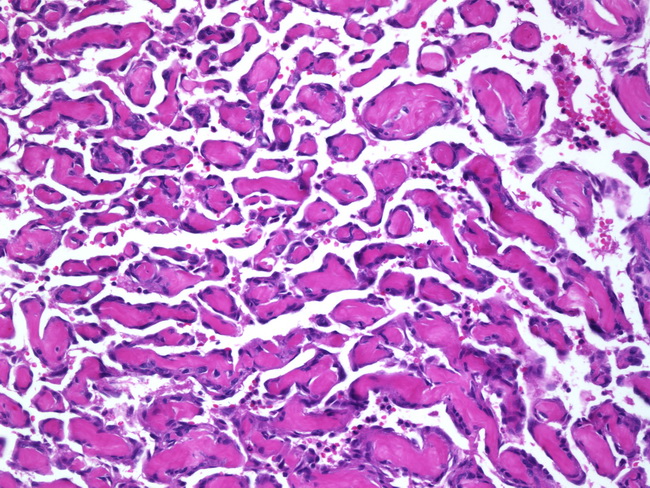
This image shows a well-established lesion from a case of papillary endothelial hyperplasia. There are numerous small delicate papillae projecting into the lumen. They consist of plump endothelium lining a collagenized core. Taken out of context, it may be mistaken for angiosarcoma. However, one should remember that angiosarcomas are never confined to a vascular lumen.
slide 3 of 3