
Embryonal Rhabdomyosarcoma : Paratesticular


Comments:
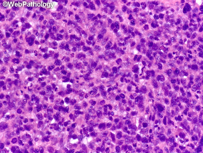
The image shows paratesticular embryonal rhabdomyosarcoma (RMS) in a 7 y/o male. RMS is the most common sarcoma of the paratesticular region in children. The peak incidence is seen around 9 years of age. It may arise in testicular tunics, epididymis, or spermatic cord. Testis proper is usually spared. In bulky tumors, the exact site of origin cannot be determined. Most paratesticular RMS are embryonal type and consist of small, round cells with hyperchromatic nuclei and scant cytoplasm giving them a basophilic appearance at low magnification. Mitotic activity is brisk. Rhabdomyoblasts are scant or absent. Rare cases have alveolar, botryoid, or pleomorphic appearance. Treatment of Paratesticular RMS: Treatment usually consists of radical inguinal orchiectomy with high ligation of the spermatic cord and ipsilateral or bilateral retroperitoneal lymph node dissection (RPLND). If imaging studies rule out involvement of retroperitoneal lymph nodes, then RPLND is not necessary. With adjuvant radiation therapy and combination chemotherapy along with surgery, the survival rates for paratesticular RMS are greater than 80%.