
Proliferative Myositis : Differential


Comments:
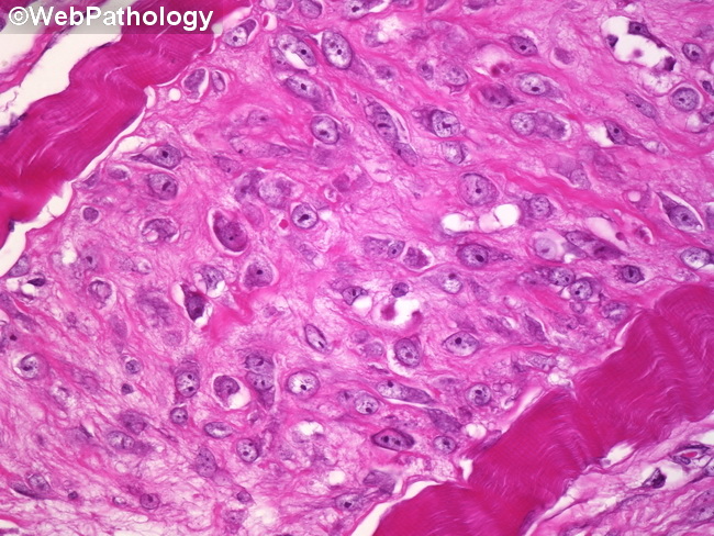
Differential Diagnosis: Proliferative myositis can mimic embryonal rhabdomyosarcoma and ganglioneuroblastoma, especially in the pediatric age group, given the presence of large ganglion-like atypical cells. Many cases are diagnosed as some type of sarcoma in children. Features favoring proliferative myositis include rapid growth, short duration of lesion, maximum size of under 3 cm (in most cases), and absence of fibrillary background. The atypical myofibroblasts of proliferative myositis lack cross striations and are basophilic (in contrast to dense eosinophilia in rhabdomyoblasts). Desmin is usually positive in proliferative myositis; however, MyoD1 and myogenin are negative. This high power image shows ganglion-like cells in proliferative myositis.