
Proliferative Myositis : Microscopic


Comments:
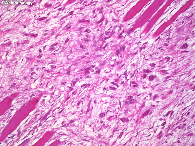
Microscopically, proliferative myositis consists of proliferation of fibroblasts and myofibroblasts in a background of myxoid stroma. This process tends to permeate the skeletal muscle rather than completely replacing large areas of the involved muscle. The skeletal muscle fibers surrounded by fibroblastic proliferation may undergo secondary atrophy. A characteristic finding in proliferative myositis is the presence of large basophilic cells resembling ganglion cells or rhabdomyoblasts. They have enlarged vesicular nuclei with prominent nucleoli. Ultrastructurally, they have features of myofibroblasts. There is increased mitotic activity; however, atypical mitoses are not found. Areas of metaplastic bone or cartilage may be present.