
Myolipoma : Microscopic Features

slide 6 of 12


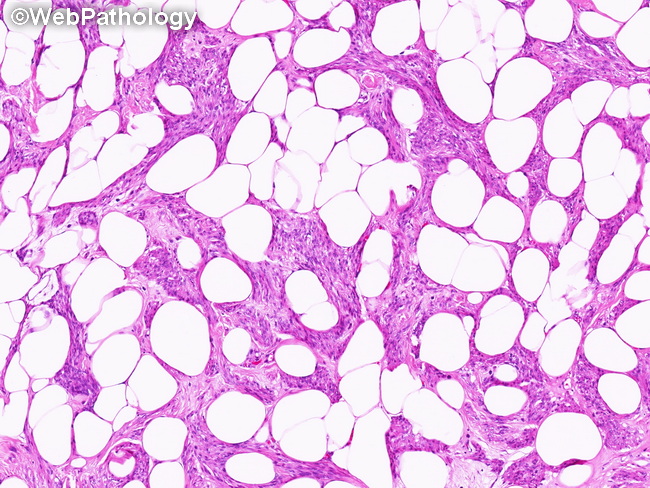
Comments:
Microscopic Features (continued from the previous image): The image shows bundles of bland smooth muscle cells separated by mature adipocytes. Variable amounts of hyalinized or edematous stroma containing mast cells are present in most cases. Uncommon features include focal hypercellularity, degenerative nuclear atypia, round cell morphology, stromal sclerosis, lymphocytic aggregates, chondroid or osseous metaplasia and hemosiderin deposition.
slide 6 of 12