
Parosteal Osteosarcoma

slide 71 of 93


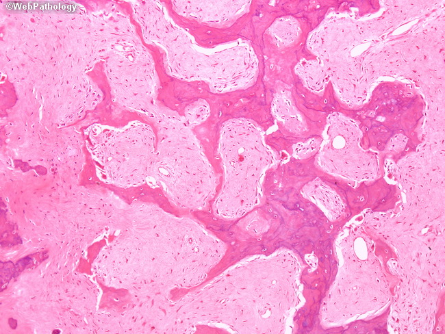
Comments:
Parosteal osteosarcoma consists of regularly arranged parallel or anastomosing bony trabeculae (shown here) separated by a hypocellular spindle stroma lacking significant atypia. There is abundant collagen deposition between spindle tumor cells which show only occasional mitotic figures. At the periphery of the lesion, the stroma is more cellular and osteoid matrix is scant. The spindle cells may infiltrate the surrounding skeletal muscle and soft tissues.
slide 71 of 93