
Long-segment Barrett Esophagus

slide 5 of 36


Comments:
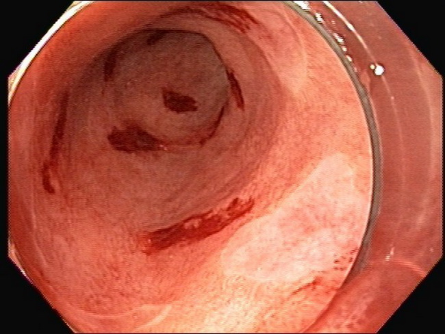
Here's another endoscopic image of long-segment Barrett esophagus (metaplastic epithelium extends at least 3 cm above the esophagogastric junction). Most of the distal esophagus is lined by metaplastic columnar epithelium. The small lighter patches are the only areas lined by squamous epithelium. The red hemorrhagic areas mark the biopsy sites. Narrow band imaging endoscopy highlights the columnar- and squamous-lined areas more clearly and is shown in the next image. Image courtesy of: Pramod Malik, MD; used with permission.
slide 5 of 36