
Allergic Fungal Sinusitis : Pathology
Home
 Infectious Disease
Filamentous Fungi (Molds)
Dematiaceous Molds (Phaeohyphomycosis)
Allergic Fungal Sinusitis : Pathology
Infectious Disease
Filamentous Fungi (Molds)
Dematiaceous Molds (Phaeohyphomycosis)
Allergic Fungal Sinusitis : Pathology

slide 2 of 8


Comments:
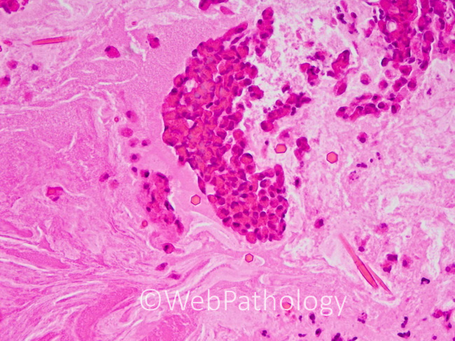
Allergic fungal sinusitis: Macroscopically, the specimen removed from the sinus is thick, rubbery, and gelatinous with a greenish-brown color. Microscopically, it consists of "allergic mucin" which appears amorphous and chondroid-like with clusters of inflammatory cells. It may have a laminated appearance. The inflammatory cells show a predominance of eosinophils and scattered neutrophils, plasma cells, and histiocytes. Charcot-Leyden crystals and desquamated respiratory cells are also frequently found. This image shows rare fungal hyphae on the upper left and lower right.
slide 2 of 8