
BK/Polyomavirus Nephritis

Comments:
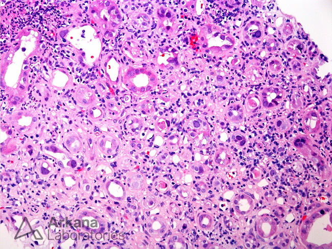
This H&E photomicrograph shows a kidney biopsy from a renal transplant recipient. There is interstitial inflammation and tubular atrophy. Within tubules, multiple epithelial cells show basophilic intranuclear inclusions and smudged chromatin. These findings, in the setting of renal transplant, are consistent with BK/Polyomavirus nephritis. An SV40 stain was performed and showed positivity within numerous tubular nuclei throughout the sample confirming BK/Polyomavirus nephritis. Image courtesy of: Arkana Labs.; used with permission.