
Melanoma Immunohistochemistry : HMB-45


Comments:
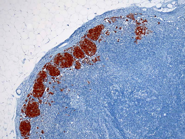
Immunohistochemical stains play a vital role in the diagnosis of amelanotic, spindle cell, and epithelioid variants of melanoma and their distinction from poorly-differentiated carcinomas as well as mesenchymal tumors. A classic case of melanoma is immunoreactive for S-100 protein, HMB-45, Melan-A, tyrosinase, Microphthalmia Transcription Factor (MITF), and vimentin. HMB-45 is less sensitive but more specific than S-100 protein as a melanoma marker. It is positive in 80-86% of metastatic deposits and in 90-100% of primary melanomas. It is quite useful in the diagnosis of amelanotic melanomas. The staining pattern is more often focal than with S-100 protein. Desmoplastic melanomas are generally negative for HMB-45. Common nevi may be HMB-45 positive in their superficial and junctional components but are negative in deeper aspects. Spitz nevus shows HMB-45 positivity in its superficial portion but is negative in deeper aspects. In contrast, spitzoid melanoma is HMB-45 positive throughout the tumor. HMB-45 also stains PEComas (angiomyolipoma, lymphangiomyomatosis, and clear cell sugar tumor of lung). The image shows metastatic melanoma infiltrates in the marginal sinus of a lymph node. Tumor cells are strongly positive for HMB-45. Image copyright: pathorama.ch.