
Neurofibromatosis 2 : Multiple Meningiomas

slide 3 of 10


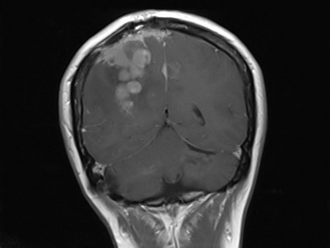
Comments:
Same case as the previous image. The MRI (Coronal post-contrast T1 image) clearly shows moderately enhancing dural based extra-axial masses in interhemispheric fissure in frontal and high parietal regions attached to the falx cerebri, indicative of multiple meningiomas. The patient also had bilateral vestibular schwannomas and was diagnosed with neurofibromatosis 2. Case courtesy of Dr Imran Ahmad Khan, Radiopaedia.org. From the case rID: 42833
slide 3 of 10