Apr 2017
Schwannoma
Reviewer(s): Dharam M. Ramnani, MD
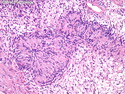
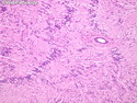
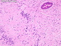
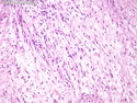
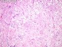
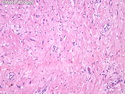
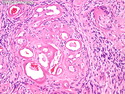
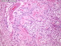
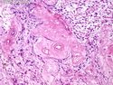
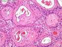
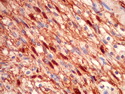
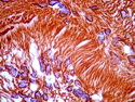
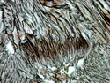
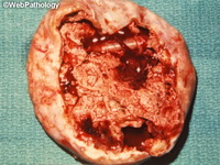
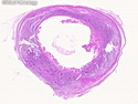
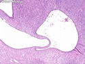
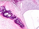
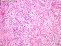
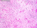
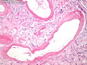
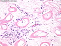
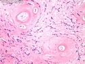
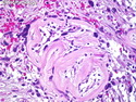
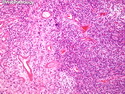
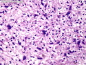
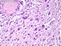
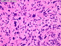
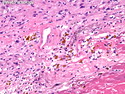
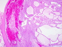
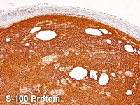
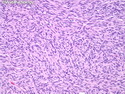
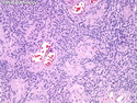
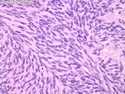
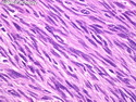
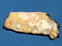
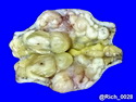
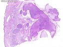
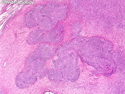
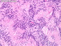
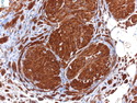
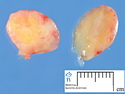
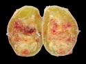
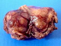
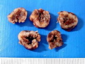
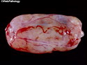
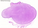
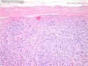
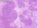
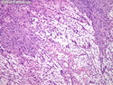
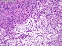
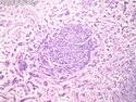
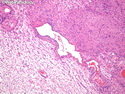
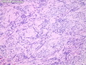
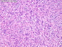
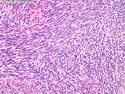
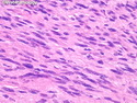
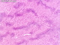
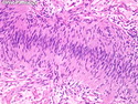
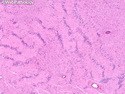
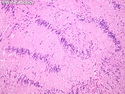
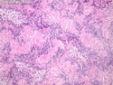
Schwannoma is a slow-growing, encapsulated benign peripheral nerve sheath tumor that is usually seen sporadically (90% of cases). The remainder are associated with Neurofibromatosis 2 (3%), schwannomatosis (2%), or multiple meningiomas with or without NF2 (5%). Schwannomas may rarely arise in the setting of NF1. The most common locations are head, neck, upper and lower extremities, posterior mediastinum, and retroperitoneum. The cut surface is yellow-white or gray. Secondary changes such as hemorrhage, cystic change, cytologic atypia, and calcification are more frequent in the larger deep-seated tumors. Schwannomas are composed of cellular Antoni A areas alternating with hypocellular Antoni B areas. Schwann cells in Antoni A areas show nuclear palisading, whorling patterns, and Verocay bodies. Antoni B areas consist of spindle cells in a loose myxoid or hyalinized stroma, inflammatory cells, collagen fibers, and ectatic deformed vessels with hyalinized walls and thrombi. Schwann cells consistently show positivity for S-100 and SOX10. Cellular schwannoma is composed of mainly Antoni A areas and shows increased mitotic activity (usually < 4 mitoses/10 HPF). They are usually found in deep-seated locations and behave in a benign fashion. Plexiform schwannomas are superficial and multinodular and make up about 5% of all schwannomas. The linkage to Neurofibromatosis 1 or 2 is weak, unlike the strong association between plexiform neurofibroma and NF1. References: 1) Enzinger & Weiss's Soft Tissue Tumors, Sixth Edition, 2014; p. 813-829.
.jpg)