Heterotopic Ossification
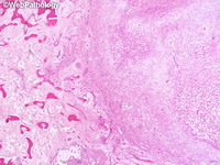
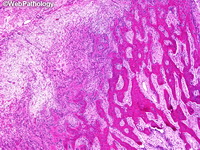
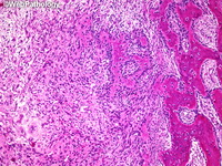
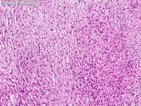
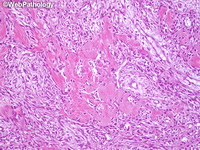
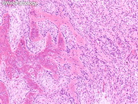
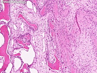
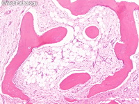
Myositis ossificans (MO) is a benign, solitary, self-limiting, ossifying mass found within the musculature of the extremities. It is related to fibro-osseous pseudotumor of digits and soft tissue aneurysmal bone cyst. Similar lesions within subcutaneous fat or tendons/fascia are referred to as panniculitis ossificans and fasciitis ossificans respectively It is cured by simple excision and has excellent prognosis. MO occurs in physically active adolescents or young adults with a male predominance and is usually associated with trauma. Skeletal muscles of thigh, buttock, shoulder and elbow are often involved. The onset is rapid (in 1-2 weeks) with pain/tenderness, soft tissue swelling and edema at the site of trauma. It goes through 3 parallel stages of clinical, radiologic and histopathologic changes (zonal pattern). The initial stage (1-4 weeks) shows highly cellular fibroblastic/myofibroblastic proliferation that can mimic extraskeletal osteosarcoma. Calcification appears in intermediate stage (4-8 weeks). The mature stage (> 8 weeks) shows abundant mature lamellar bone that can mimic osteoma. There is controversy regarding any malignant potential of MO. Grossly, mature lesions have a hard peripheral shell of ossification and a soft, gelatinous, grey-red center. The pathogenesis underlying inappropriate differentiation of fibroblasts into osteoblasts with bone formation is poorly understood. Some cases show USP6 rearrangements. The differential diagnosis includes benign and malignant conditions including: extraskeletal osteosarcoma, parosteal osteosarcoma, metastasis (osteoblastic carcinoma, melanoma) and benign reactive processes such as nodular fasciitis, proliferative myositis, and exuberant fracture callus. Additional images and a more detailed discussion are under Myositis Ossificans in Orthopedic Pathology section.