Jan 2016
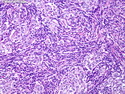
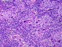
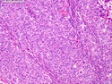
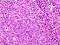
Medullary Carcinoma
Reviewer(s): Dharam M. Ramnani, MD
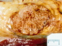
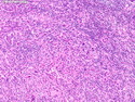
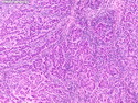
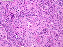
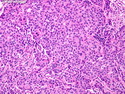
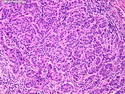
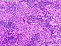
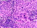
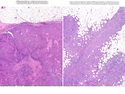
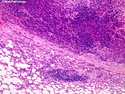
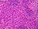
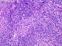
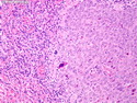
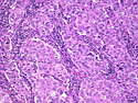
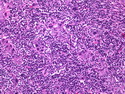
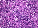
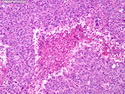
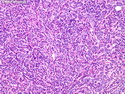
Medullary carcinomas make up less than 5% of all breast carcinomas. They share a number of features with BRCA-1 associated breast cancers, including - relatively young age at diagnosis, lympho-plasmacytic infiltrate, high-grade morphology, triple-negative phenotype, and p53 mutations. Among breast cancers arising in BRCA-1 carriers, about 13% are medullary carcinomas. They are bulky, soft, and well-circumscribed with pushing borders that don�t infiltrate into the surrounding breast tissue or fat. They show diffuse growth pattern with high-grade nuclei, frequent mitoses and abundant eosinophilic cytoplasm with a syncytial appearance. There is a prominent lymphoplasmacytic infiltrate within and around the tumor composed of peripheral T-cells (with numerous activated cytotoxic lymphocytes) and IgA-producing plasma cells. It is believed to be host reaction to the tumor and is thought to play a role in better prognosis seen with this tumor as compared to other breast cancers. The 10-yr survival rate is about 84%. The immunohistochemical profile of medullary carcinoma is similar to conventional invasive ductal carcinonma. They are usually positive for CK7, S-100 protein, and P53. Cytokeratin 20 is generally negative. ER, PR, and HER2 are negative (triple negative phenotype).
.jpg)