Microglandular Adenosis
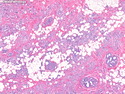
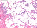
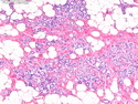
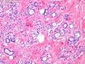
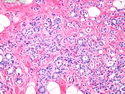
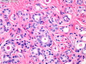
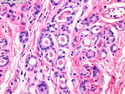
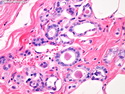
Microglandular adenosis (MGA) is an uncommon proliferative lesion that mimics carcinoma clinically and morphologically. It is composed of small uniform glands irregularly distributed in fibrofatty stroma. The glands are lined by a single layer of uniform cuboidal or flattened cells with regular nuclei and punctuate nucleoli. The glandular lumens are round and contain eosinophilic secretions. The glands lack myoepithelial layer; however, a thick basement membrane is present that can be highlighted by reticulin stain. The main differential diagnosis is with tubular carcinoma and sclerosing adenosis. Tubular carcinoma has angular glands arranged in stellate or radial configuration. Tubular carcinoma lacks both myoepithelial cells and basement membrane and expresses ER and PR. Microglandular adenosis has a significant premalignant potential. In-situ and invasive carcinoma have been reported arising within MGA in about 30% of cases. When needle core biopsy is suggestive of MGA, the lesion should be completely excised with negative margins.