Adenocarcinoma of Prostate
High Quality Pathology Images of Genitourinary: Prostate of Adenocarcinoma of Prostate
Home
Sections
Neuropath
Glial Tumors
Non-Glial Tumors
Non-Neoplastic
Breast
Benign Prolif. Lesions
Breast Carcinomas
Rare Breast Tumors
Inflammatory/Reactive
Head & Neck
Oral Cavity, Oropharynx & Neck
Maxillofacial
Salivary Glands
Nose & Sinuses
Larynx
Ear
Eye
Mediastinum
Mediastinum
Peritoneum
Peritoneum, Omentum & Mesentery
Genitourinary
Prostate
Urinary Bladder
Kidney
Testis
External Genitalia
Adrenal
Cancer Genomics
Soft Tissue Tumors
Hematopathology
Mature B-cell Neoplasms - Part I
Mature B-cell Neoplasms - Part II
Mature T-cell & NK-cell Neoplasms
Myeloid, Histiocytic & Dendritic Cell Neoplasms
Lymph Node (Non-Hematopoietic)
Spleen
Gynecologic
Vulva/Vagina
Cervix
Uterus
Ovary
Placenta & Trophoblastic Lesions
Orthopedic
Bone Tumors - I
Bone Tumors - II
Tumor-like Lesions of Bone
Misc. Bone Lesions
Lesions of Joints
DermPath
Proliferations & Neoplasms
Non-Neoplastic DermPath - I
Non-Neoplastic DermPath - II
Endocrine
Thyroid
Parathyroid
Pancreas
Adrenal
Pituitary
Gastrointestinal
Esophagus
Stomach
Small Bowel
Appendix
Large Bowel
Anus
Liver
Gallbladder
Pancreas
Soft Tissue
Fibroblastic
Fibrohistiocytic
Lipomatous
Myogenic
Vascular & Lymphatic
Peripheral Nerve
Uncertain Histogenesis
Misc. Soft Tissue Lesions
Pulmonary
Lung - Non-Neoplastic
Epithelial Lung Tumors
Mesenchymal Tumors of Lung
Pleura
Miscellaneous Lung Tumors
Cardiovascular
Heart
Blood Vessels
Infectious Disease
Specific Clinical Syndromes
Gram-Positive Bacteria
Gram-Negative Bacteria - I
Gram-Negative Bacteria - II
Acid-Fast Bacilli
Yeast & Yeast-like Fungi
Filamentous Fungi (Molds)
Viruses
Parasites
Pediatric
Pediatric Pathology - I
Pediatric Pathology - II
Cytopathology
Cytopathology 1
Cytopathology 2
Genetic Disorders
About
Feedback
Contact
Striving to be the most comprehensive online resource for high-quality pathology images
13,153
Images
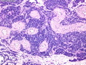
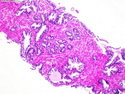
Adenocarcinoma of Prostate
Reviewer(s): Dharam M. Ramnani, M.D.
Home
Genitourinary
Prostate
Adenocarcinoma of Prostate
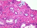
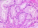
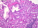
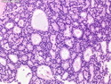
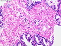
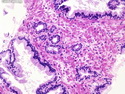
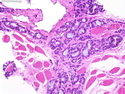
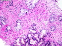
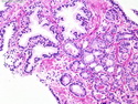
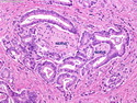
Adenocarcinoma of Prostate : Histologic Features
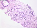
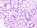
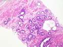
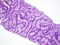
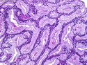
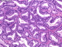
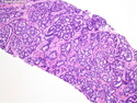
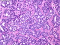
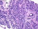
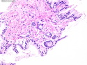
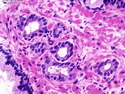
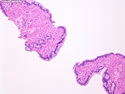
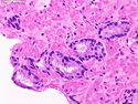
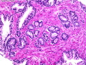
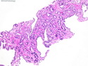
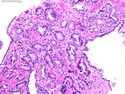
Adenocarcinoma of Prostate : Architecture
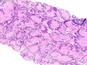
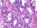
Adenocarcinoma of Prostate : Architecture
Adenocarcinoma of Prostate : Architecture
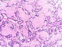
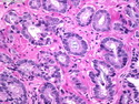
Adenocarcinoma of Prostate : Architecture
Adenocarcinoma of Prostate : Architecture
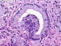
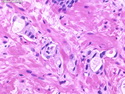
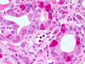
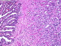
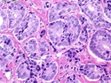
Adenocarcinoma of Prostate : Cytologic Features
Adenocarcinoma of Prostate : Cytologic Features
Adenocarcinoma of Prostate : Cytologic Features
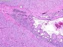
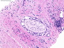
Adenocarcinoma of Prostate : Perineural Invasion
Adenocarcinoma of Prostate : Perineural Invasion
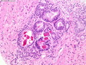
Adenocarcinoma of Prostate : Crystalloids
Adenocarcinoma of Prostate : Blue Mucin
Adenocarcinoma of Prostate : Blue Mucin
Adenocarcinoma of Prostate : Collagenous Micronodules
Adenocarcinoma of Prostate : Collagenous Micronodules
Adenocarcinoma of Prostate : Glomerulations
Gleason Grading System
Gleason Grading System : Overview
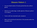
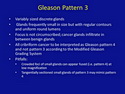
Gleason Pattern 1
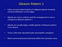
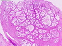
Gleason Pattern 2
Gleason Pattern 2
Gleason Pattern 2
Gleason Score 3+2
Gleason Score 3+2
Gleason Score 3+2
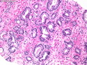
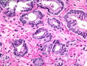
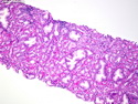
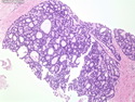
Gleason Pattern 3
Gleason Score 3+3
Gleason Score 3+3
Gleason Score 3+3
Gleason Score 3+3
Gleason Score 3+3
Gleason Score 3+3
Gleason Score 3+3
Gleason Score 3+3
Gleason Score 3+3
Gleason Score 3+3
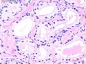
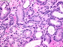
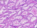
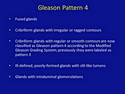
Gleason Pattern 4
Gleason Score 4+4
Gleason Score 4+4
Gleason Score 4+4; Hypernephromatoid
Gleason Score 4+4
Gleason Score 4+4
Gleason Score 4+4
Gleason Score 4+4
Gleason Score 4+4
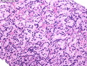
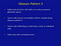
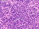
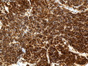
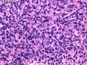
Gleason Pattern 5
Gleason Score 5+5
Gleason Score 5+5; PSA Immunostain
Gleason Score 5+5
Gleason Score 5+5
Gleason Score 5+5
Gleason Score 5+5
Gleason Score 5+5; Comedonecrosis
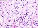
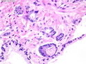
Minimal Prostate Cancer
Minimal Prostate Cancer
Minimal Prostate Cancer
Minimal Prostate Cancer
Minimal Prostate Cancer
Minimal Prostate Cancer
Minimal Prostate Cancer
Minimal Prostate Cancer
Minimal Prostate Cancer
Minimal Prostate Cancer
Minimal Prostate Cancer : Racemase
Minimal Prostate Cancer
Minimal Prostate Cancer : HMWCK + p63
Prostate Cancer : Miscellaneous Features
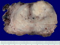
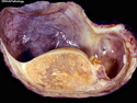
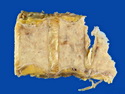
Prostate Cancer Gross Specimen
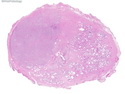
Prostate Cancer : Whole Mount
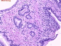
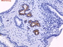
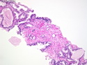
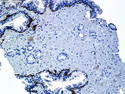
Neuroendocrine Cells in Prostate Cancer
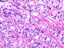
Prostate Cancer in Skeletal Muscle
Prostate Cancer in Seminal Vesicle
Prostate Cancer in Seminal Vesicle
Prostate Cancer in Seminal Vesicle
Prostate Cancer : Testicular Metastases
Prostate Cancer : Testicular Metastases
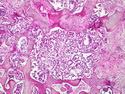
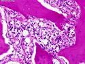
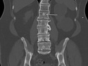
Prostate Cancer : Bone Metastases
Prostate Cancer : Bone Metastases
Prostate Cancer : Bone Metastases
Prostate Cancer : Bone Metastases
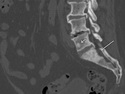
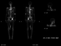
Prostate Cancer : Bone Scan
Prostate Cancer : Bone Metastases
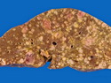
Prostate Cancer : Liver Metastases

.jpg)